| Back | Seborrhoeic Keratosis |
| Appearance |
A seborrheic keratosis is a benign skin growth that often develops as we get older and it has been estimated that over 90% of adults over the age of
60 years have one or more of them. They occur in males and females of all races, typically beginning to erupt in the 30s or 40s.
They are uncommon under the age of 20 years.They are also referred to as : ■ SK ■ Basal cell papilloma ■ Senile wart ■ Brown wart ■ Wisdom wart ■ Barnacle. Typical appearance characteristics are : |
■ A round or oval-shaped waxy or rough bump ■ Typically found on the face, chest, a shoulder or the back ■ Usually brown, black or light tan in colour ■ They appear waxy, scaly, and slightly raised. ■ Variable in size from very small to more than 2.5cm across ■ Can be single or multiple |
| Cause |
Experts don't completely understand what causes a seborrheic keratosis.
This type of skin growth does though tend to run in families, so there is likely an inherited tendency and if you've had one seborrheic keratosis, you're at risk of developing others.
A seborrheic keratosis isn't contagious or cancerous. Possible causes are : |
■ They can develop following sunburn or dermatitis. ■ Skin friction may be the reason they appear in body folds. ■ Viral cause (eg human papillomavirus) is unlikely. ■ Seborrhoeic keratosis can arise from solar lentigo. ■ Location suggests a role of UV radiation. ■ Some cancer treatments can result in warty keratoses. |
| Diagnosis |
Normally the diagnosis of seborrhoeic keratosis is often straight forwards appearing as a stuck-on, well-demarcated warty plaque.
Sometimes though, seborrhoeic keratosis may resemble skin cancer, such as basal cell carcinoma, squamous cell carcinoma or melanoma.
There are diagnostic dermatoscopic clues to seborrhoeic keratosis, such as multiple orange or brown clods (due to keratin in skin surface crevices),
white milia-like clods, and curved thick ridges and furrows forming a brain-like or cerebriform pattern. If doubt remains a partial shave, punch biopsy or diagnostic excision can be undertaken. |
| Complications |
Seborrhoeic keratoses are not premalignant tumours however sometimes they are difficult to tell apart from skin cancers.
Skin cancer may also, by chance, arise within or collide with a seborrhoeic keratosis so care has to be taken when carrying out an assessment. Very rarely, eruptive seborrhoeic keratoses (which is an irritated seborrhoeic keratosis that appears as an inflamed, red and crusted lesion) may denote an underlying internal malignancy, most often gastric adenocarcinoma. Eruptive and irritated seborrhoeic keratoses may also arise as an adverse reaction to a medication, such as adalimumab, vemurafenib, dabrafenib, 5-fluorouracil and many chemotherapy drugs. It may give rise to eczematous dermatitis around the seborrhoeic keratosis. Dermatitis may also trigger new seborrhoeic keratoses to appear. There are also several variants of seborrhoeic keratoses which include: Solar lentigo : This is a harmless patch of darkened skin often called an "old age spot" or "senile freckle" but can be difficult to distinguish from a flat seborrhoeic keratosis. They appear as pigmented patches in sun-exposed sites. Dermatosis papulosa nigra : Small, pedunculated and heavily pigmented seborrhoeic keratoses on head and neck of darker-skinned individuals. Stucco keratoses : : Grey, white or yellow papules on the lower extremities Lichenoid keratosis : : An inflammatory phase preceding involution of some seborrhoeic keratoses and solar lentigines. |
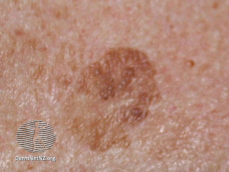 | |
| Seborrhoeic Keratosis |
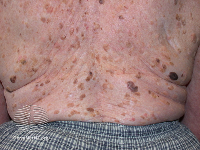 | |
| Seborrhoeic Keratosis |
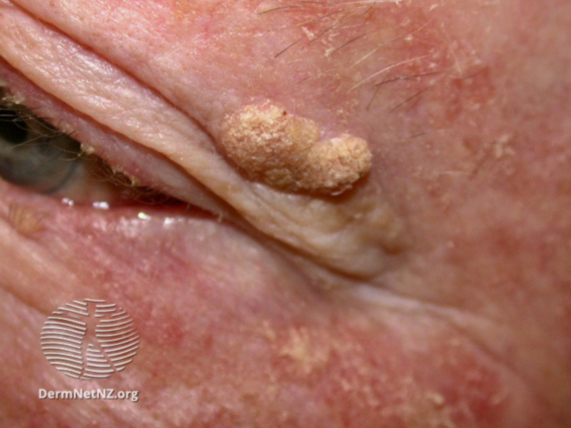 | |
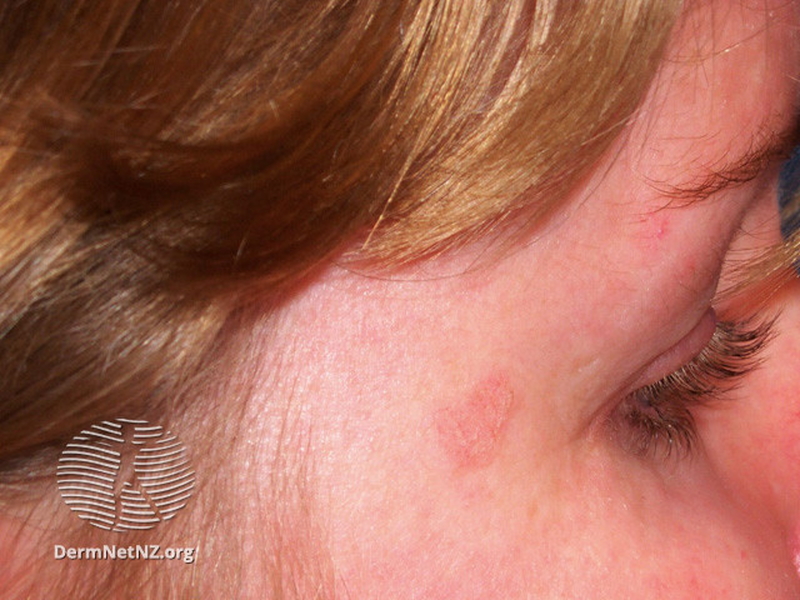
| Seborrhoeic Keratosis on the eye |
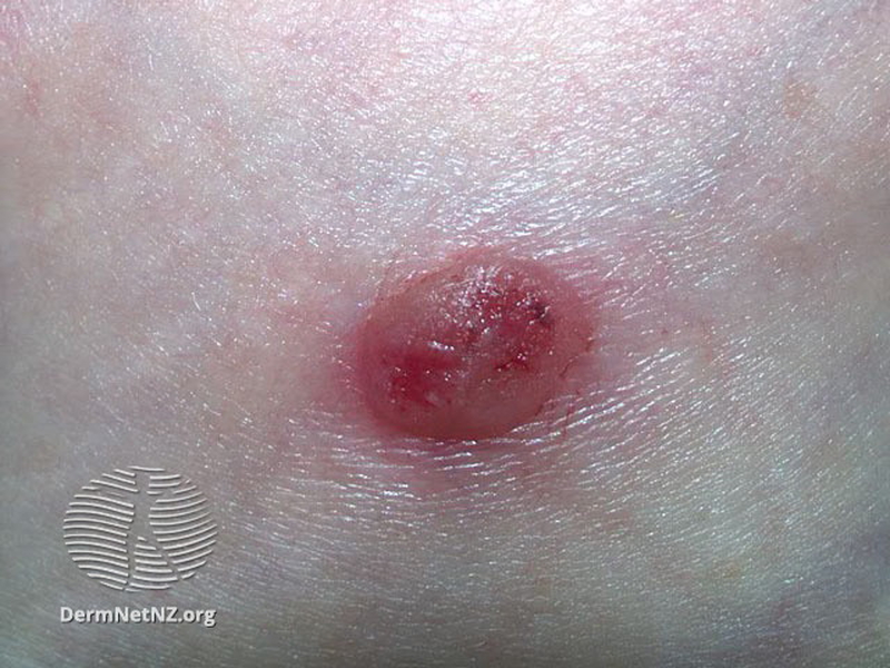 | |
| Irritated Seborrhoeic Keratosis |
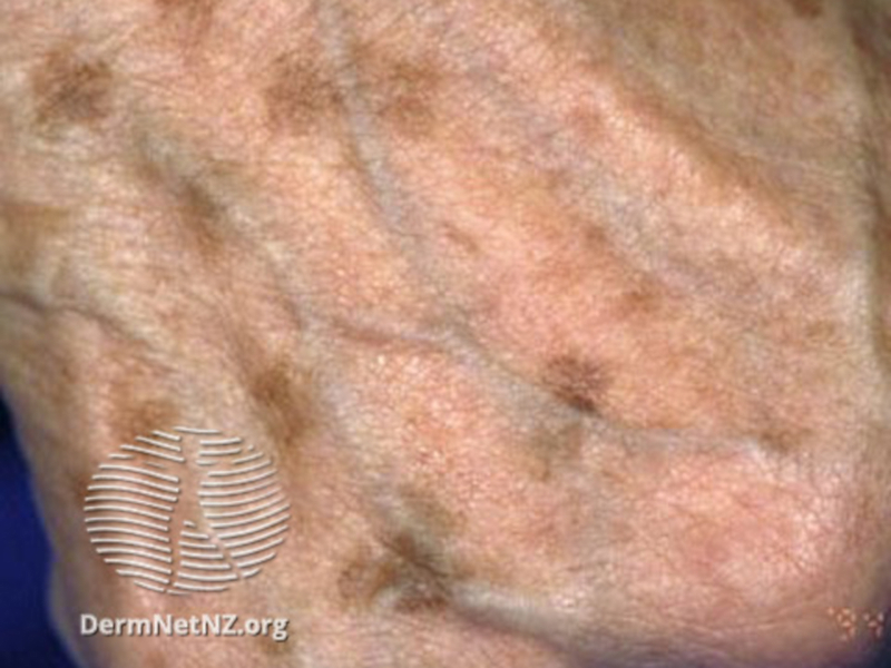 | |
| Solar Lentigo |
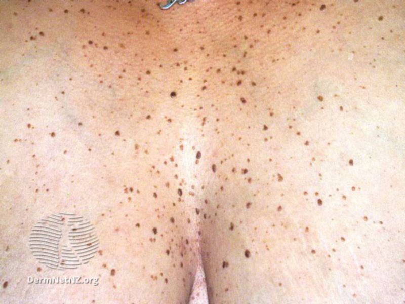 | |
| Dermatosis Papulosa Nigra |
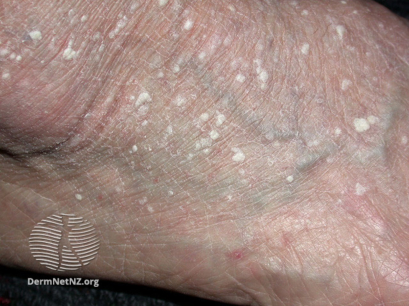 | |
| Stucco Keratosis |
 | |
| Lichenoid Keratosis |
| Treatment |
Seborrhoeic keratoses are harmless and not contagious.
They don't normally need treatment, but can be removed if they become irritated by clothing or their appearance becomes unsightly.
An individual seborrhoeic keratosis can easily be removed if desired.
Methods used to remove seborrhoeic keratoses include: Cryotherapy This uses liquid nitrogen and is suitable for thinner lesions and can be repeated if necessary. Curettage and/or Electrocautery Ablative laser surgery Shave biopsy This involves shaving off the lesion with a scalpel Focal Chemical Peel This process uses trichloracetic acid All methods have disadvantages. Treatment-induced loss of pigmentation is a particular issue for dark-skinned patients. There is no easy way to remove multiple lesions on a single occasion. |